I am a bit of an addict when it comes to radiology and as with most addictions, mine is probably not a healthy one. Let me start by saying I have zero training as a radiologist. However, most of the time I am actually able to identify the right body part (full disclosure: I have screwed this up before. It is a mess in there!). I have also been able to locate all of my tumors without someone showing me where they are. Sometimes I locate problems that no one else has pointed out yet (including the radiologist that gave the official read on the scan in question). And I think I found a problem on an MRI of my spine.
Over the course of the last 13 months, I have had really horrible nocturnal back pain. The pain is very localized, way down in my sacrum/tailbone area. I am almost 100% pain-free during the day, but nights are bad. I can barely roll over in bed, I often need to get on my hands and knees and sort of back out of bed if I do have to get up in the middle of the night. Once I am in position I can rest ok, but if I move, the pain is really bad. Once I get out of bed in the morning and move around a little bit, it seems to go away for most of the day. I do notice that if I have to lay flat on a hard surface during the day, the pain will come back too (I know this because I spend a fair amount of time laying in MRI/CT/PET scan machines). I have probably had 20 pain-free nights since it first started. I can’t talk about it without getting all teary because I am really sick of it.
I also have high bone turnover markers (which I have mentioned before), which we measure regularly by some combination of Alkaline Phosphatase, Bone-specific Alkaline Phosphatase, and N-telopeptides. We have been looking into several explanations for why this is the case over past few months. The Wizard recently ordered a bone scan to see if anything turned up, but it came back as normal. Which, as is all too common for me these days, is reassuring and annoying at the same time. I don’t want to be loaded with “hot spots” on a bone scan, but it would be nice for a scan to point to an actual explanation for the labs and my pain too! We do know that the pain can’t be something like a disc, because it is too far down in my spine for that. So, what the heck is going on down there?
Since I now have an extensive collection of images of my entire body, I decided to flip through my library and take a look myself to see if I could spot anything. I pulled up an MRI of my sacrum from June 2014 and started digging around. And I found something that looked weird to me. On the image, it looks like two black round spots in my sacrum, right about where my back was hurting:
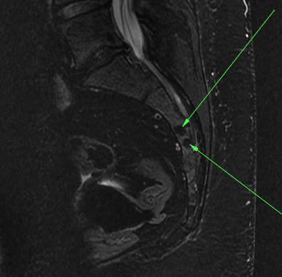
I have become pretty handy with the medical imaging software and even added the little green arrows so you can quickly spot the stuff I am concerned about. I don’t think those things are supposed to be there. I am not 100% sure, as I am not a doctor, but this doesn’t look normal to me.
So today, I went in for my first real appointment with my new PCP. I fired my last PCP recently, after we had this exchange, and I was worried that bringing this up at my first appointment with my new PCP might really give him the impression I was a lunatic. So I decided to ease into the topic and just see how it went before springing the pictures on him. Let me just say that my initial impression of this doctor is very good. He seems very engaged and eager to figure out and explain things. I chose him specifically because he was highly rated, a very experienced internal medicine doctor (not “just” a PCP), and because he chose to enter a practice where he had fewer patients and could spend more time with each of them. For a complicated patient like me, all of these attributes seemed important. My initial impression from our “meet and greet” was that he was also the kind of physician that did not mind patients that were pro-active and had a lot of questions and wanted to understand the details of medical problems and the treatments. Also important to me.
We talked about the fact that I was having surgery next week. He knows my surgeons and is going to touch base with them and stop by and see me post-op in the hospital too. He certainly doesn’t have to do that but I REALLY appreciate that he is going to do that. He had lots of suggestions for things I should be asking anesthesia tomorrow during my final pre-op appointment and I really appreciated that as well. So then we got into my real agenda for the day – this bone pain. I showed him my bone labs from the Wizard and told him I had a normal bone scan. I explained the pain to him and the fact that it tended to only show up at night. I brought in the CD of my spine MRI as well and asked if he wanted to take a look. Unfortunately, he couldn’t load it on his computer right there in the office.
So….I paused and told him that I had looked at it myself and I thought I saw something. I know, doctor, I am not a radiologist, but I took a screen shot of my MRI and I have it right here on my iPhone. Want to take a look? I cringed inside as I asked him (OMG he is going to think I am a lunatic, OMG I am a lunatic). But to my surprise, he said “Sure, let me have a look!” After a scramble to dig it out, I showed him the above image and…he agreed with me! That’s not normal! Then he went a step further. He pulled out an anatomy book and gave me an education on my sacrum! I could not have been happier.
First he showed me a picture of what the sacrum looks like from the back:
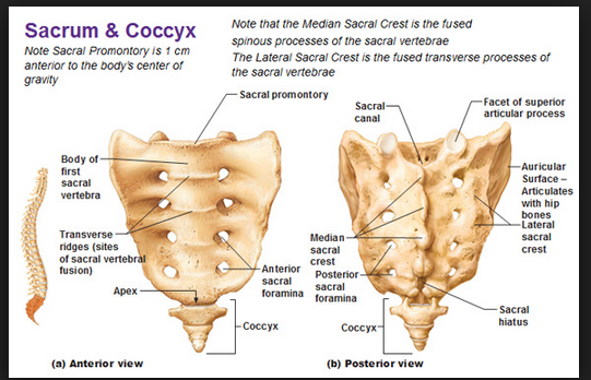
So, you can see the sacrum does have two rows of holes in it. Those holes, the sacral foramina, are where the nerves run through the sacrum. I had actually seen these holes before when I was educating myself on the sacrum when trying to figure out if that is what I was seeing, just from a weird angle. So, I was glad to have him walk through this with me.
Then he pulled up a picture of my sacrum from the side. I will put the picture of my MRI below for comparison purposes:
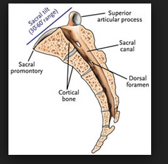
The main point, if you compare these images, is that the black circles in my MRI are not in the same location as the foramina. The sacral canal, where the foramina are located, is above and to the right of my black circles. Comparing the same two MRI views that match the anatomy pictures, you can the black circles are in a different location than the foramina:

The green box shows the black circles from the back view (the left image) and the side view (the right image). The foramina can be seen in the left hand side view. It is the larger dark circle to the right of the green box, as indicated by the green arrow. Those black circles in the green box aren’t supposed to be there.
The next question of course is what the hell are those things then? The short answer is that we don’t know yet. My current theory is that my high bone turnover markers, my pain and this MRI all mean bone tumors. I would be lying if I said bone metastasis hadn’t crossed my mind – nocturnal bone pain like mine and the bone turnover markers would be consistent with that. And NETs like the one we are planning to evict next week often spread to lungs, liver and bone (and I have “stuff” in all those locations). I am not saying that to be melodramatic or cause anyone to lose sleep, me included. But the whole point of this blog was just for me to lay out what is going on and what I am thinking. And this is what I am thinking. It could be nothing (that’s not likely though, let’s be honest). It could be something. I really don’t know. All I know is that this shit needs to get sorted out one way or the other.
And my new favorite PCP agrees – this needs further investigation. So, the plan is to see where we are at when the dust settles after surgery next week. And after the dust settles, he is ordering a new MRI of the spine (as this one is already a year old) and he is sending me to a highly regarded bone doctor. He is starting to get that referral in place now, in case there is a long wait. As usual, I am half worried and half relieved that someone is listening to me and taking this stuff seriously.
Let me tell you the other thing that made me want to run around my new PCP’s desk and hug him. When I described my bone pain to him, he questioned me about it then concluded: “That is so strange that your bones only hurt at night. It makes no sense to me. But you aren’t crazy, and if you say it only hurts at night, it only hurts at night. I just can’t give you an explanation for WHY that is the case.” AHHHH THANK YOU!! Thank you for just listening to what I am experiencing and believing me. Thank you for not telling me that my experiences can’t be right or that I must be experiencing something else. It sounds so obvious that this should be the way things actually work in doctor-patient relationships. But so often it doesn’t.
The big question you might have on your mind right now is the following: you had this MRI a year ago because of bone pain. What did the radiology report say? Didn’t it mention anything? Well, here is what happened last year. PCP #1 (I am now on PCP #3) ordered the MRI when I told her about my bone pain in May 2014. Here is what she emailed to me. This is a verbatim cut and paste from the summary of the radiology report.
 IMPRESSION:
     No evidence of sacral fracture.

     L5-S1 degenerative disc disease as above.
OK, that’s good news. My sacrum isn’t broken. I knew I had disc problems too, so that wasn’t a surprise. But since the MRI didn’t appear to provide any information that would be useful in helping us sort out my bone pain, I decided I would just go and look at the report myself. And here is what the rest of the report actually said.
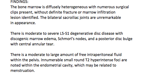
OK, now that is just weird. The report is claiming I have “numerous surgical clips present” but to my knowledge, I have never had back surgery. The report seems to be saying further that there is no evidence to tell them how the clips got in there. Is it just me or does this not make sense? It seems kind of clear to me at this point that my PCP either never read the full report OR she didn’t find it puzzling that I was full of surgical clips and complaining about back pain and never mentioned a prior back surgery to her. So I emailed her to say “what’s up with this report and can you please have the radiologist look at this again because this doesn’t make any sense.” So she did. The radiologist in turn corrected my report so that it now says:
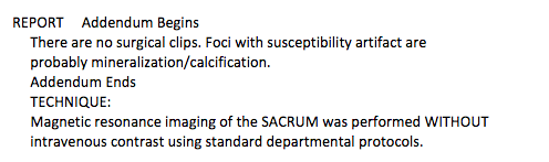
OK. So my back is NOT full of surgical clips. This is good news to me, as I could take “alien abduction and resulting back surgery” off the differential diagnosis list. Now I’m told it is “probably mineralization.” And I don’t really know what that means or if it is harmless or significant and no one bothers to explain it to me. I have never managed to actually locate anything that looks like a surgical clip in my MRI, for the record, so I am still not even sure where this mineralization is. All I have been able to locate is those black circles. But it is really disturbing to me that the initial report was so bizarre, that this MRI was read TWICE because the patient pointed out the report didn’t make any sense, and despite all of this no one managed to see or comment on what appears to be a problem so obvious an economist can spot it on the MRI.
The moral of the story dear reader: AUDIT EVERYTHING. And don’t stop digging until you know what is wrong with you. I am mad at myself for not pushing someone harder to look at the MRI for a THIRD time LAST YEAR. I am mad at myself for not sitting down with my own CD of the MRI and making sure it was normal. In my defense, I was sick and this was one of many problems I was trying to figure out. And it was also clear my PCP was getting fed up with me for pestering her with this when it was clear she believed there was nothing wrong beyond old age and back pain that goes with that territory. AND IT WAS THE RADIOLOGIST’S JOB TO READ THE SCAN PROPERLY, NOT MINE. But what is done is done. All I can do now is keep pestering anyone who will listen – and fire everyone who won’t.
